
分享至
简介
基本信息
技术文档
上下游信息
相关文章
评价
咨询
售后
产品简介
最大吸收波长/发射波长: 748/780 nm 推荐滤光片设置: 710 ex/760 em 图1.从脾脏分离的T细胞用DiR和i.v进行荧光染色。 (5x10⁶个细胞/小鼠)注射到Nu / Nu小鼠中。 上面使用IVIS Spectrum注射后24小时拍摄的图像显示,细胞归巢于脾脏。 DiR染料是亲脂性的近红外花菁荧光染料,可以用来染细胞膜和其它脂溶性生物结构。18个碳的长链插入细胞膜中从而对细胞进行染色, 而细胞间的染料转移可以忽略不计。DiR的发射的是近红外荧光可以穿透细胞和组织,在活体成像中用来示踪。 DIR一般对原代细胞进行荧光染色并可进行活体成像分布观察, (例如下列细胞embryonic stem cells, bone marrow derived stem cells, adipose derived stem cells, lymphocytes and erythrocytes), 图2.通过将25 mg溶于3 mL乙醇来制备DiR储备液。 通过在5 mL PBS中稀释199 µL储备液来制备320 µg / mL的工作溶液。 从脾脏分离的T细胞与320 µg / mL DiR孵育。 孵育30分钟后,将细胞在4ºC下以1000 rpm离心离心3分钟,得到蓝色沉淀。 将细胞在PBS中洗涤两次并静脉内注射(5×10⁶细胞/小鼠)。 对照组在PBS中每只小鼠注射5×10⁶个细胞。 在注射后10分钟,1小时,6小时和24小时用IVIS Spectrum对小鼠成像。 用于DiR成像的理想滤光片组是710 nm激发和760 nm发射。 在所有时间点对小鼠的背侧和腹侧成像。 注射后24小时,收集脑,骨头,脾,肝,肺和肾用于离体成像。 非侵入性体内成像实时显示了向肝脏和脾脏注射的T细胞的归巢过程,这通过离体成像得到了证实。 参考文献 Kalchenko et al., Use of lipophilic near-infrared dye in whole-body optical imaging of hematopoietic cell homing. Journal of Biomedical optics, September/October 2006, Vol 11(5). Product Introduction Maximum absorption wavelength/emission wavelength: 748/780 nm Recommended filter setting: 710 ex/760 em Figure 1. T-cells isolated from the spleen were fluorescently stained with DiR and i.v. injected (5x10⁶ cells/mouse) into a Nu/Nu mouse. Images above taken 24hrs post injection with IVIS Spectrum show cells homing to the spleen. DiR dye is a lipophilic near-infrared cyanine fluorescent dye that can be used to dye cell membranes and other fat-soluble biological structures. A long chain of 18 carbons is inserted into the cell membrane to stain cells, while the transfer of dye between cells is negligible. DiR emits near-infrared fluorescence that can penetrate cells and tissues and is used for tracking in in vivo imaging. DIR generally performs fluorescent staining of primary cells and can observe the distribution of in vivo imaging, (For example, the following cell embryonic stem cells, bone marrow derived stem cells, adipose derived stem cells, lymphocytes and erythrocytes), Figure 2. DiR stock was prepared by dissolving 25 mg in 3 mL ethanol. Working solution of 320 µg/mL was prepared by diluting 199 µL of stock solution in 5 mL PBS. T-cells isolated from the spleen were incubated with 320 µg/mL DiR. After 30 min incubation, cells were spun down for 3 min at 1000 rpm at 4 ºC resulting in a blue pellet. Cells were washed twice in PBS and injected intravenously (5 x 10⁶ cells/mouse). Control group was injected with 5 x 10⁶ cells/mouse in PBS. Mice were imaged with IVIS Spectrum at 10 min, 1hr, 6hr and 24 hrs post injection. Ideal filter set for DiR imaging is 710 nm excitation and 760 nm emission. Mice were imaged dorsally as well as ventrally at all time points. Brain, bones, spleen, liver, lungs and kidneys were harvested for ex vivo imaging 24 hrs post injection. Non-invasive in vivo imaging showed the homing process of injected T cells to the liver and spleen in real time, which was confirmed by ex vivo imaging. References Kalchenko et al., Use of lipophilic near-infrared dye in whole-body optical imaging of hematopoietic cell homing. Journal of Biomedical optics, September/October 2006, Vol 11(5).
中文名称
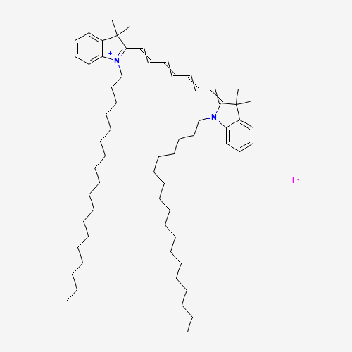
DiR' [DiIC18(7)],用于膜染色
英文名称
DiR' [DiIC18(7)]
中文别名
2-(7-(3,3-二甲基-1-十八烷基吲哚啉-2-亚基)庚-1,3,5-三烯-1-基)-3,3-二甲基-1-十八烷基-3H-吲哚-1-鎓碘化物
英文别名
2-(7-(3,3-Dimethyl-1-octadecylindolin-2-ylidene)hepta-1,3,5-trien-1-yl)-3,3-dimethyl-1-octadecyl-3H-indol-1-ium iodide
CAS号
100068-60-8
分子式
C63H101IN2
分子量
1013.41 g/mol
精确质量
≥95%
PSA
-
Logp
-
技术文档
「暂无技术文档」
上下游信息
「暂无上下游信息」
相关文章
「暂无相关文章」
用户评价
加载中...
商品咨询

温馨提示:

因厂家可能临时调整产品规格、价格、库存、参数等信息且不另行通知,同时每位用户的咨询时间、采购需求存在差异,平台回复内容仅在一定时期内具有参考价值。由此给您带来的不便,敬请谅解,感谢您的理解与支持!最新详细信息请咨询在线客服,一切以客服实时回复为准!

售后服务

收到货品请及时核对数量、外观及产品信息,如遇破损、错发、质量异常等问题,欢迎随时联系我们。

服务热线:400-6226-992 ,我们将第一时间为您处理,感谢您的支持与信任!

DiR' [DiIC18(7)] 98% 10mg

DiR' [DiIC18(7)] 98% 10mg 麦克林 100068-60-8,规格齐全,生产科研实验检测,科学材料数智化一站式易购平台。

品牌:麦克林
货号:D909616-10mg
级别: -
货期:30天
已售 553 件
976
.8
1074
.5
配送至: 请选择地址
运费:0 元
数量

精选好物